
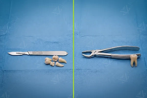
Tooth extraction is sometimes the best option for preserving your overall oral health. Whether a tooth is severely damaged, impacted, or compromising neighbouring teeth, removing it can prevent further complications and open the door to restorative solutions. Understanding the difference between simple and surgical extraction helps you prepare for the procedure, set realistic expectations for recovery, and feel confident in the treatment plan your dentist recommends.
Simple (Non-Surgical) Extraction
A simple extraction is performed on teeth that are visible and fully erupted in the mouth. The tooth can be seen above the gumline, and its roots follow a relatively straightforward anatomy. This is the most common type of extraction and is routinely performed by general dentists as well as oral surgeons.
How a Simple Extraction Works
The procedure is straightforward. After administering local anaesthesia to numb the area completely, the dentist uses two primary instruments:
- An elevator -- a lever-like instrument placed between the tooth and the surrounding bone to loosen the tooth from its periodontal ligament
- Forceps -- used to grip the loosened tooth and remove it with controlled rocking and rotational movements
A simple extraction typically takes under 30 minutes, often considerably less. The socket is checked for any remaining fragments, and gauze is placed to control bleeding. In most cases, sutures are not required.
When Simple Extraction Is Appropriate
- Severely decayed teeth that cannot be restored with a filling, crown, or root canal treatment
- Loose teeth from advanced gum disease (periodontitis) where the supporting bone has been significantly lost
- Orthodontic treatment -- strategic extractions to create space for proper alignment of remaining teeth
- Baby teeth that have not fallen out naturally and are blocking the eruption of permanent teeth
- Supernumerary (extra) teeth that are causing crowding or other complications
Surgical Extraction
A surgical extraction is required when a tooth is not easily accessible through the mouth. This may be because the tooth has not fully erupted through the gum, has broken off at or below the gumline, or has root anatomy that prevents straightforward removal. Surgical extractions are typically performed by oral and maxillofacial surgeons or dentists with advanced surgical training.
What Makes It "Surgical"
Unlike a simple extraction, a surgical procedure involves one or more of the following additional steps:
- Incision in the gum tissue -- a flap of gum tissue is raised to expose the tooth and surrounding bone
- Bone removal -- a small amount of bone covering the tooth may need to be removed using a surgical handpiece
- Tooth sectioning -- the tooth is divided into two or more pieces, allowing each section to be removed individually through a smaller opening
- Suturing -- the gum tissue is repositioned and sutured closed after the extraction
More complex anaesthesia options may be available for surgical extractions, including conscious sedation (oral or intravenous) for patients with dental anxiety or for lengthy procedures.
When Surgical Extraction Is Needed
- Impacted wisdom teeth -- fully impacted (completely buried in bone) or partially impacted (partially visible through the gum). This is the most common indication for surgical extraction
- Broken teeth at the gumline -- when a tooth has fractured and insufficient crown structure remains above the gum for forceps to grip
- Teeth with curved, long, or divergent roots -- unusual root anatomy that prevents the tooth from being removed in one piece through the socket
- Teeth with dense surrounding bone -- particularly common in the lower jaw, where thick cortical bone resists conventional extraction forces
- Ankylosed teeth -- teeth that have fused directly to the bone, eliminating the normal periodontal ligament space. These teeth cannot be loosened with an elevator and must be sectioned from the bone surgically
The Surgical Extraction Procedure: Step by Step
1. Imaging and Planning
Before any surgical extraction, detailed imaging is essential. At Vision Dental Clinic, we use CBCT (Cone Beam Computed Tomography) 3D scans to visualise the tooth in three dimensions. This reveals the exact position of the tooth, the number and curvature of its roots, proximity to important anatomical structures (such as the inferior alveolar nerve in the lower jaw or the maxillary sinus in the upper jaw), and the density and volume of surrounding bone.
A CBCT scan transforms a surgical extraction from an exercise in uncertainty to a precisely planned procedure. Every critical structure is mapped before a single incision is made.
2. Anaesthesia
Local anaesthesia is administered to completely numb the tooth and surrounding tissues. For complex cases or anxious patients, conscious sedation may be offered in addition to local anaesthesia. The area is thoroughly tested for numbness before the procedure begins.
3. Incision and Flap Elevation
A carefully planned incision is made in the gum tissue, and a mucoperiosteal flap is raised to expose the tooth and surrounding bone. The incision design is chosen to provide optimal access while ensuring good blood supply to the tissue for healing.
4. Bone Removal (If Required)
When bone covers the tooth or prevents its path of removal, a surgical handpiece with a round bur is used under copious irrigation to remove the necessary amount of bone. Only the minimum bone needed for access is removed.
5. Tooth Sectioning
For multi-rooted teeth or teeth with complex anatomy, the tooth is divided into sections using a surgical bur. Each section is then removed individually, requiring less force and a smaller bony opening than removing the entire tooth in one piece.
6. Extraction
The tooth (or tooth sections) is removed using elevators and forceps. The socket is thoroughly inspected and debrided to ensure no root fragments, bone spicules, or infected tissue remains.
7. Site Cleaning and Suturing
The extraction site is irrigated with sterile saline to remove debris. If bone grafting is planned for future implant placement, the graft material is placed at this stage. The gum flap is repositioned and secured with sutures, which may be resorbable (dissolve on their own) or non-resorbable (removed at a follow-up appointment).
Recovery: Simple vs Surgical Extraction
Simple Extraction Recovery (1 to 3 Days)
Most patients experience only mild discomfort after a simple extraction, peaking within the first 24 hours and resolving within 2 to 3 days. Over-the-counter pain medication is usually sufficient. Light bleeding and minor swelling are normal. Most patients return to normal activities the following day.
Surgical Extraction Recovery (5 to 7 Days)
Surgical extractions involve more tissue manipulation and therefore require a longer recovery period. Swelling typically peaks at 48 to 72 hours and gradually subsides over the following days. Prescription pain medication may be provided for the first 2 to 3 days. Most patients feel significantly better by day 5 and return to full activities within a week.
Post-Operative Care Instructions
Regardless of the type of extraction, following these guidelines promotes optimal healing:
- Gauze pressure -- bite firmly on the gauze pad for 30 to 45 minutes after the procedure. Replace with a fresh pad if bleeding continues
- Ice packs -- apply to the outside of the cheek for 20 minutes on, 20 minutes off during the first 24 hours to minimise swelling
- Soft diet -- eat soft foods (yoghurt, soup, mashed potatoes, smoothies) for the first 2 to 3 days. Avoid hot foods and drinks for the first 24 hours
- Pain management -- take prescribed or recommended pain medication as directed. Do not wait for pain to become severe before taking medication
- Avoid straws and smoking -- the sucking action can dislodge the blood clot and cause dry socket. Avoid straws for at least 5 days and smoking for at least 72 hours (ideally longer)
- Oral hygiene -- brush teeth normally but avoid the extraction site for 24 hours. After 24 hours, gently rinse with warm salt water (half a teaspoon of salt in a glass of warm water) 3 to 4 times daily
- Rest -- avoid strenuous physical activity for 48 to 72 hours after surgical extractions to prevent increased bleeding
Potential Complications and How to Avoid Them
Dry Socket (Alveolar Osteitis)
Dry socket occurs when the blood clot in the extraction site is dislodged or dissolves before healing is complete, exposing the underlying bone to air, food, and bacteria. It causes significant pain that typically begins 2 to 4 days after extraction and may radiate to the ear. Dry socket is more common after lower wisdom tooth extractions and in patients who smoke. Treatment involves gentle irrigation of the socket and placement of a medicated dressing to relieve pain and promote healing.
Infection
Post-extraction infection is uncommon but can occur, particularly when pre-existing infection was present. Signs include increasing pain, swelling, fever, and pus discharge from the site. Antibiotics may be prescribed preventatively for patients with compromised immune systems or when extracting heavily infected teeth.
Nerve Damage (Rare)
The inferior alveolar nerve runs through the lower jaw near the roots of the lower wisdom teeth. In rare cases, surgical extraction of impacted lower wisdom teeth can cause temporary or (very rarely) permanent numbness or altered sensation in the lower lip, chin, or tongue. CBCT imaging allows precise assessment of the nerve's proximity to the tooth roots, enabling the surgeon to plan accordingly and minimise this risk.
Sinus Communication (Upper Teeth)
The roots of upper premolars and molars sit close to the floor of the maxillary sinus. Occasionally, extraction of these teeth creates a small communication between the mouth and the sinus cavity. If this occurs, it is identified immediately and managed with appropriate closure techniques. Patients are instructed to avoid blowing their nose forcefully and to sneeze with their mouth open for several days to allow the site to heal.
Dr. Hussien's Oral Surgery Expertise
At Vision Dental Clinic Abu Dhabi, surgical extractions are performed by Dr. Hussien Tahoun, who brings over 35 years of experience in oral and maxillofacial surgery. Dr. Hussien has performed thousands of surgical extractions, from routine wisdom tooth removals to complex cases involving deeply impacted teeth, ankylosed teeth, and teeth in close proximity to vital structures.
Every surgical extraction at Vision Dental Clinic begins with a comprehensive CBCT scan and a thorough discussion of the procedure, risks, and recovery expectations. Dr. Hussien's approach prioritises minimally invasive technique -- removing only the bone necessary for access, sectioning teeth strategically to reduce force, and placing sutures meticulously to promote primary healing. This philosophy results in less post-operative pain, faster recovery, and fewer complications.
When surgery is indicated, precision and experience make the difference between a straightforward recovery and unnecessary complications.