
MOHAP LicensedNo. 5889VHC5-110626
Book Appointment
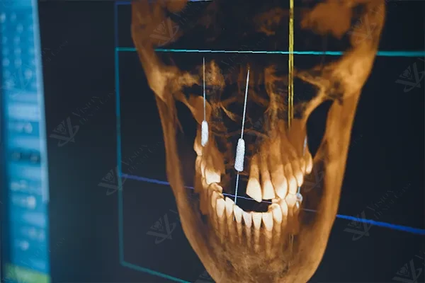
Dental surgery has entered a new era of precision and safety, driven largely by advances in imaging technology. At the heart of this transformation is the CBCT scan, a powerful diagnostic tool that gives dentists and oral surgeons an unprecedented three-dimensional view of the structures inside your jaw. Whether you are considering dental implants, facing a wisdom tooth extraction, or undergoing any procedure that involves the bone and soft tissues of the mouth, a CBCT scan has become an essential part of the planning process.
CBCT stands for Cone Beam Computed Tomography. It is a specialised type of X-ray technology that uses a cone-shaped beam of radiation to capture a complete three-dimensional image of your teeth, jawbone, nerve pathways, and surrounding soft tissues in a single rotation around your head. The scanner produces hundreds of individual images during the rotation, which are then reconstructed by software into a highly detailed 3D model.
Unlike a medical CT scanner, which uses a fan-shaped beam and requires higher radiation doses, the CBCT unit is compact, designed specifically for the head and neck region, and delivers significantly lower radiation. The result is a diagnostic tool that provides extraordinary detail with minimal exposure, making it ideally suited for dental and maxillofacial applications.
Traditional dental X-rays, such as periapical or panoramic radiographs, produce flat, two-dimensional images. While these are useful for detecting cavities, assessing the general condition of teeth, and viewing the overall layout of the jaw, they have inherent limitations:
A CBCT scan overcomes all of these limitations. It produces a true 3D dataset that can be viewed from any angle, sliced in any plane, and measured with sub-millimetre accuracy. The clinician can rotate the image, zoom into specific areas, and examine structures that would be invisible or ambiguous on a conventional X-ray.
If a traditional X-ray is like looking at a shadow on a wall, a CBCT scan is like holding the actual object in your hands and examining it from every direction.
Performing surgery in the oral and maxillofacial region means working in close proximity to critical anatomical structures: nerves, blood vessels, sinuses, and varying densities of bone. A CBCT scan provides the surgeon with the detailed roadmap needed to navigate these structures safely and effectively.
The inferior alveolar nerve runs through the lower jaw and supplies sensation to the lower lip, chin, and teeth. During procedures like implant placement or wisdom tooth extraction in the lower jaw, accidental damage to this nerve can result in temporary or permanent numbness. A CBCT scan reveals the exact position and path of this nerve, allowing the surgeon to plan the procedure to maintain a safe distance from it at all times.
For dental implants to succeed, they need to be placed in bone that has adequate height, width, and density to support them. A CBCT scan provides precise measurements of available bone in all three dimensions. If the bone is insufficient, the scan helps determine whether a bone graft or sinus lift procedure is needed and how it should be performed.
Infections, cysts, tumours, and other pathological conditions are not always visible on standard X-rays, particularly when they are small or located in areas that are difficult to image in two dimensions. A CBCT scan can reveal these hidden conditions before they become complications during surgery, allowing the treatment plan to be adjusted accordingly.
The maxillary sinuses sit directly above the upper back teeth. When placing implants in this region, it is critical to know the exact position and shape of the sinus floor. A CBCT scan shows the sinus anatomy in full detail, enabling the surgeon to determine whether a sinus lift is required and to plan the procedure with precision.
Perhaps the most transformative advantage of CBCT technology is its role in digital surgical planning. Using the 3D data from the scan, the surgeon can virtually place implants on a computer screen, experimenting with different positions, angles, and implant sizes until the optimal plan is found. This virtual plan can then be used to manufacture a custom surgical guide, a physical template that fits over the teeth and directs the drill to the exact planned position during the actual surgery.
The versatility of CBCT technology means it is valuable across a wide range of dental disciplines, not just implant surgery:
This is the most common application. CBCT allows for precise assessment of the implant site, selection of the appropriate implant size, and creation of surgical guides for guided implant placement. The result is safer, faster, and more predictable implant surgery.
Impacted wisdom teeth often have roots that curve around or sit in close proximity to the inferior alveolar nerve. A CBCT scan shows the exact relationship between the tooth roots and the nerve canal, helping the surgeon decide on the safest approach for extraction.
Complex root canal anatomy, including curved canals, additional canals that are not visible on standard X-rays, and the presence of periapical lesions, can be clearly visualised with a CBCT scan. This is particularly valuable in retreatment cases or when standard radiographs do not explain persistent symptoms.
CBCT imaging can reveal impacted teeth, supernumerary teeth, and the three-dimensional relationship of teeth within the jaw. This information helps orthodontists plan more accurate and efficient tooth movement strategies.
The temporomandibular joints (TMJ) are complex structures that are difficult to assess with conventional imaging. A CBCT scan can reveal bony changes in the joint surfaces, such as erosion, flattening, or osteophyte formation, aiding in the diagnosis and management of temporomandibular disorders.
If your dentist recommends a CBCT scan, there is very little to prepare for. The experience is straightforward, quick, and entirely painless:
At Vision Dental Clinic in Abu Dhabi, CBCT imaging is an integral part of our diagnostic and surgical workflow. Our in-house CBCT scanner means that patients do not need to visit a separate imaging centre; the scan is performed on-site, and the results are reviewed immediately by the treating clinician.
Dr. Hussien Tahoun, our oral and maxillofacial surgeon with over 35 years of experience and more than 5,000 implants placed, uses CBCT data as the foundation of every implant case. Each implant is digitally planned on the 3D scan before any surgery takes place. Custom surgical guides are then produced from this digital plan, ensuring that the implant is placed at the exact position, angle, and depth that was determined to be optimal.
This CBCT-guided approach delivers measurable benefits: shorter surgical time, less post-operative discomfort, faster healing, and higher long-term success rates. It represents the standard of care that every patient considering dental surgery should expect.
At Vision Dental Clinic, no implant is placed without a CBCT scan. Digital planning is not optional; it is the foundation of every surgical case we undertake.
Keep Reading
Everything you need to know about dental implants, from the procedure to long-term care and maintenance.
Read articleUnderstand the difference between simple and surgical extractions and what to expect from each procedure.
Read articleRoot canal therapy preserves your natural tooth and eliminates infection. Learn how the procedure works.
Read articleStart Your Journey
Book a consultation at Vision Dental Clinic. Our in-house CBCT scanner provides instant 3D imaging so your treatment is planned with accuracy from day one.